Entry #1.5: Elephant Ear Stem Fiber
May 17, 2025
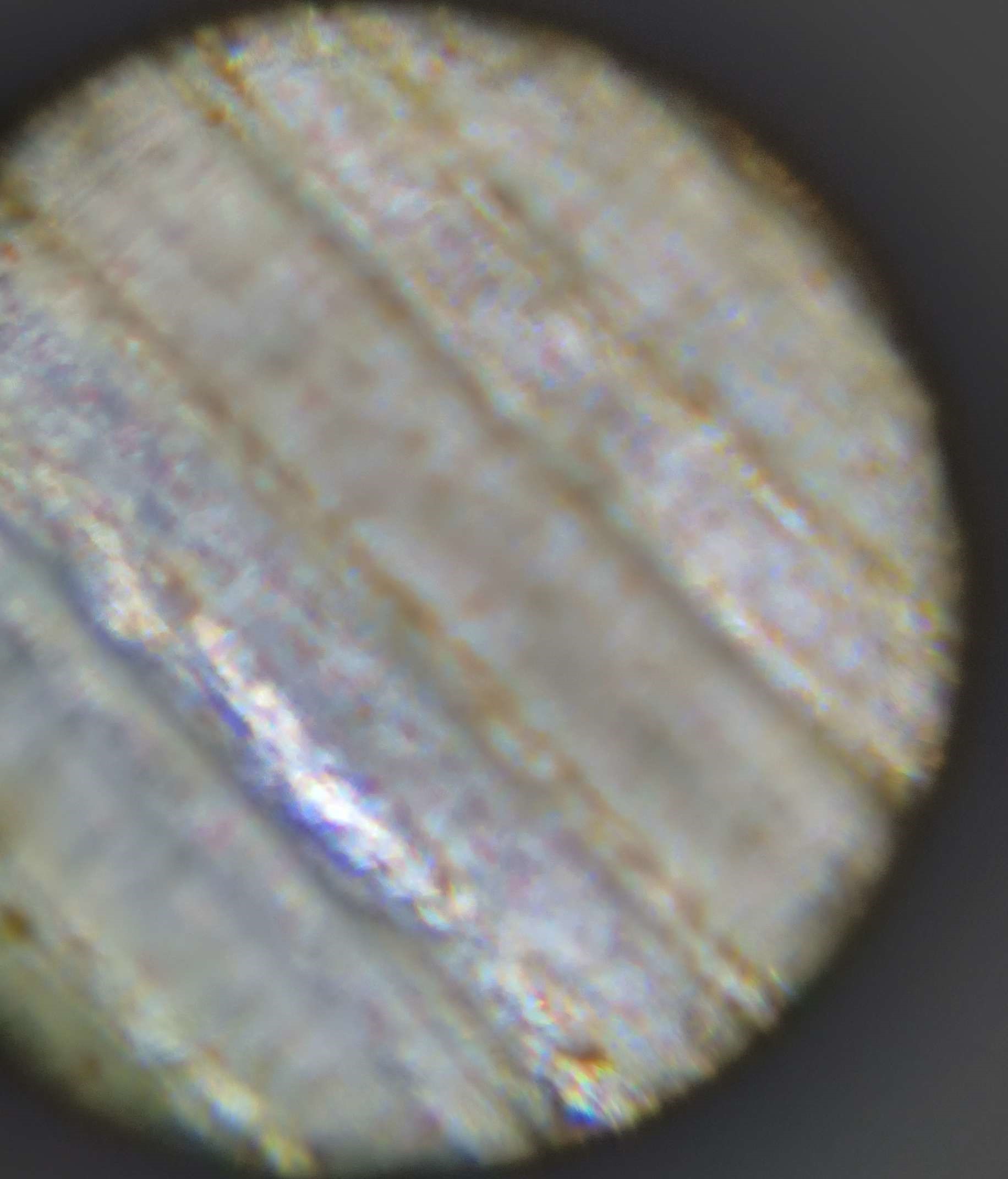
So the plant cells containing the red dot pigments (which I presumed were anthocyanins) are not actually part of the cell wall. I scraped it thin and the core was responsible for showing that image. The actual cell wall just contains longitudinal plant cells.
Entry #1: Elephant Ear Stem
June 21, 2025
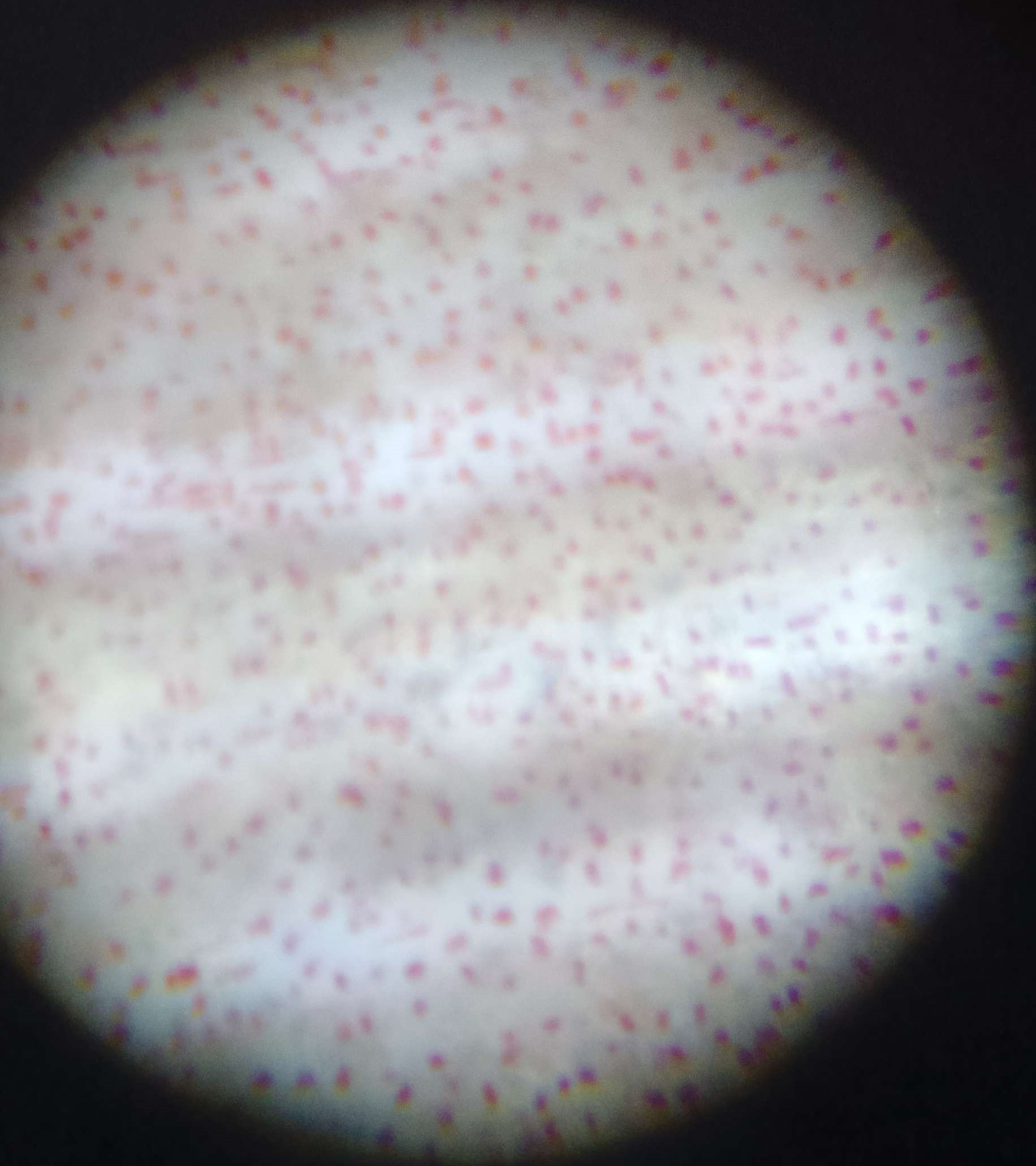
Today I collected a thin outer layer of the baby stem of an Elephant Ear plant and put it under the microscope, I saw clear long straight cell walls but focused the image to the red dots which I assumed were cells, but after asking ChatGPT (I know shameless) it says it could be anthocyanins which are red dot pigments in young leaves that protect against UV radiation and pests.